The trachea and the lungs of theBrazilian Amazon turtle, Podocnemis expansa (Testudines, Podocnemididae): morphological and ultrastructural analysis
DOI:
https://doi.org/10.65053/asa20190102Keywords:
trachea, lung, morphology, Podocnemis expansa, Brazilian amazon turtleAbstract
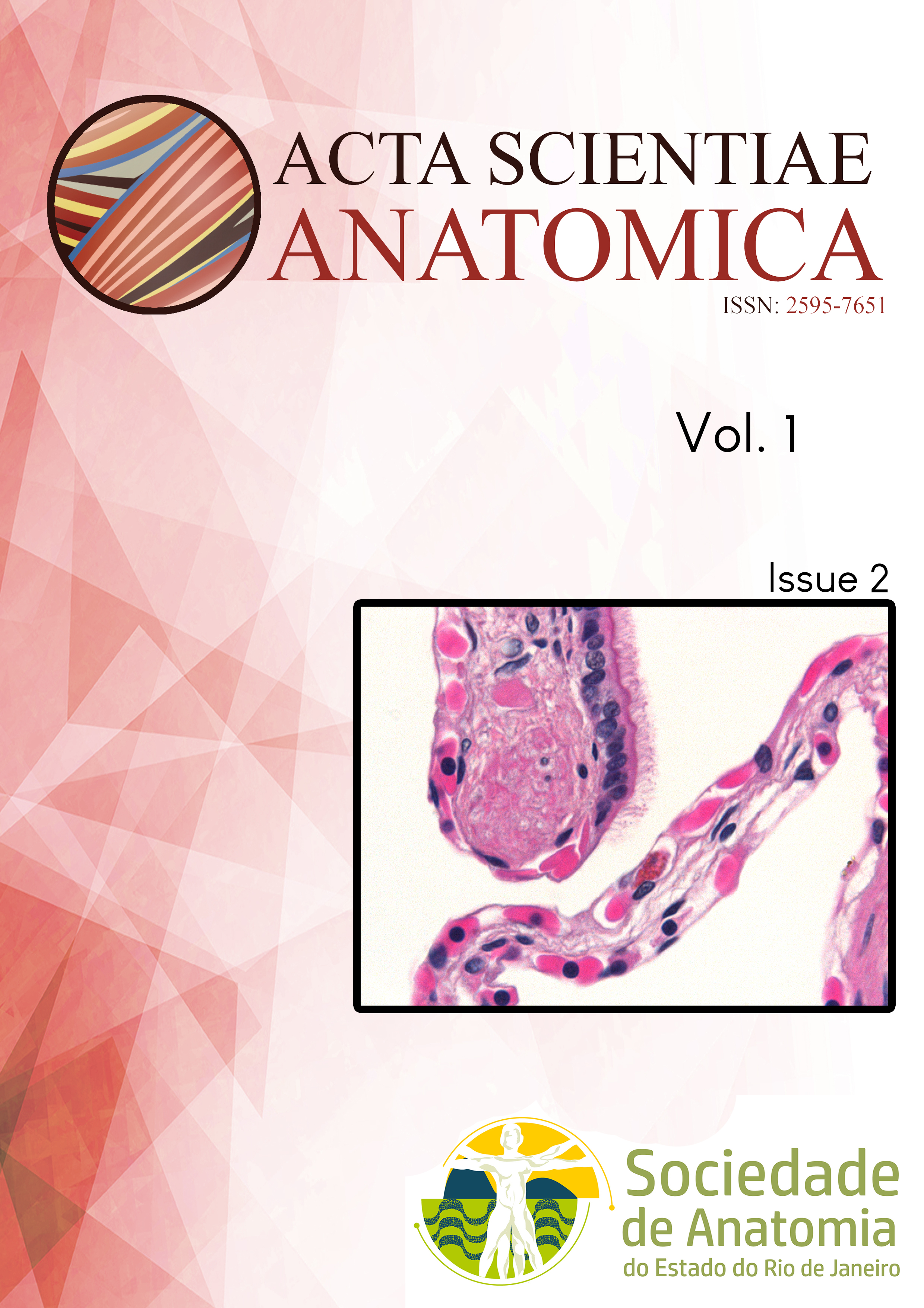
The respiratory system of the turtle is formed by specific organs that convey air from environment to small air sacs used for gas exchange called alveoli. In Podocnemis expansa, the trachea bifurcates into two primary extrapulmonary bronchi that, at the hilum, enters each lung of a spongy appearance, and are located just beneath the carapace. Four tissue layer structure the trachea: mucosa, submucosa, cartilage and serosa. The mucosa is lined by typical respiratory epithelium, resting on lamina propria of loose connective tissue. Just beneath, the submucosa is poorly developed and continuous to the perichondrium of the hyaline cartilage piece. The serosa is constituted by connective tissue with elastic fibers. The intrapulmonary bronchus exhibits similar trachea morphology but the cartilage pieces are fragmented. Inside the lung, projections depart from the pulmonary wall and project into the lumen, forming cup-like chambers named alveoli. Considering the projection size and the type of the lining epithelium, they are classified as primary, secondary and tertiary trabeculae. Two types of pneumocytes lined the lateral surface of trabeculae: lining pneumocyte type I and pneumocyte type II, which is also responsible for surfactant production. The trabeculae is supported by loose connective tissue rich in blood vessels, containing collagenous fibers, scarce elastic fibers as well as smooth muscles cells. The morphological analysis revealed that the trachea and the lung of Brazilian Amazon turtle possess similarities with other chelonian species with phylogenetic proximity.
Downloads
Published
Issue
Section
License
Copyright (c) 2025 Acta Scientiae Anatomica

This work is licensed under a Creative Commons Attribution-NonCommercial-ShareAlike 4.0 International License.
This journal publishes open-access articles under the Creative Commons Attribution 4.0 International (CC BY 4.0) license. This permits use, sharing, adaptation, distribution, and reproduction in any medium or format, as long as appropriate credit is given to the authors and the source, a link to the license is provided, and any changes are indicated. License: https://creativecommons.org/licenses/by/4.0/


