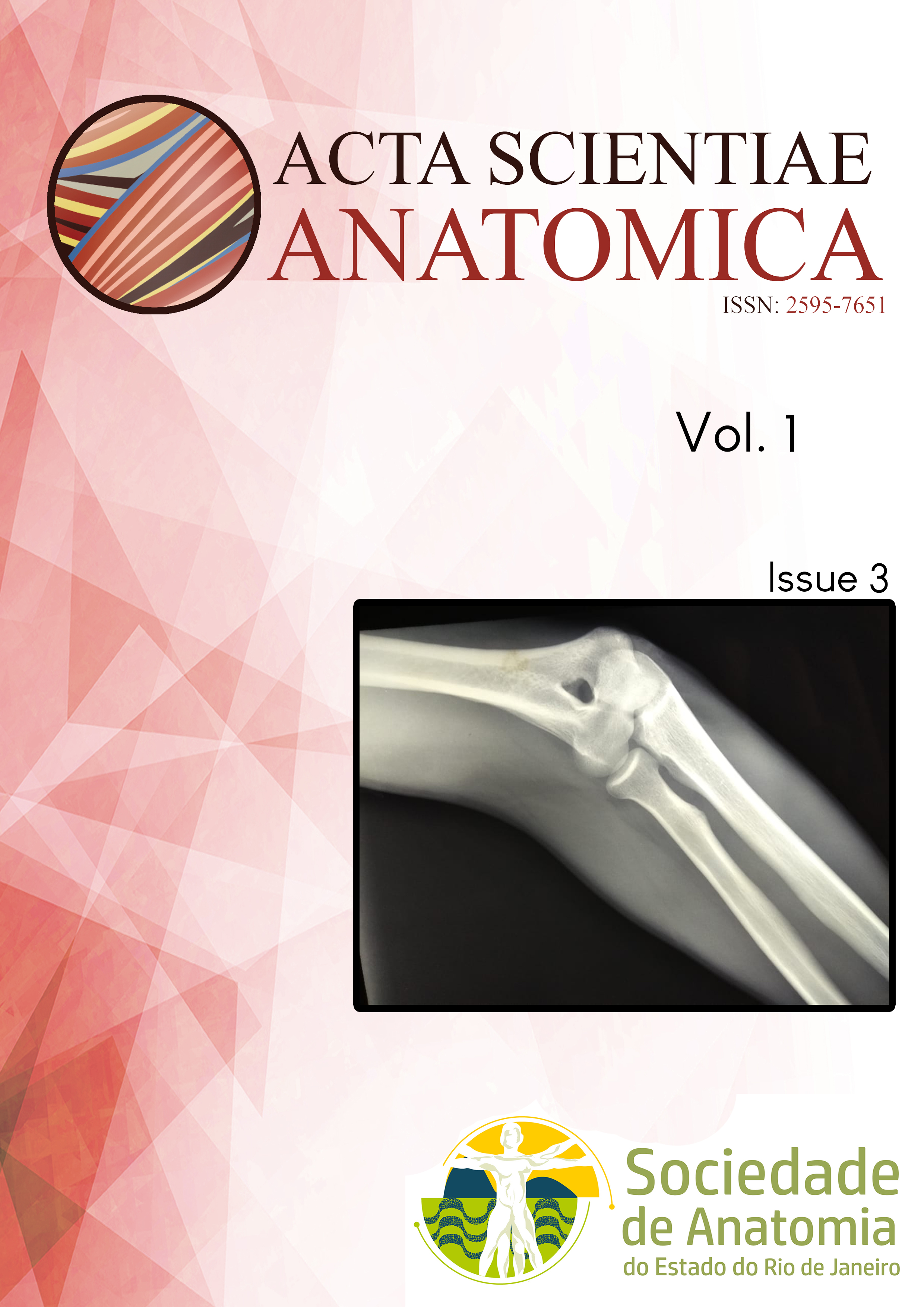
Olecranon aperture: an incidental finding
DOI:
https://doi.org/10.65053/asa20190309Keywords:
anatomy, supratrochlear foramen, humerus variation, olecranon aperture, septal apertureAbstract
The olecranon aperture or the supratrochlear foramen is an aperture situated at the distal epyphysis of the humerus. This aperture is situated between the olecranon fossae and the coronoid fossae. These fossae are usually separated by a thin bony plate or membrane. The olecranon aperture is an anatomical variant said to be present in at least 20% of the population, with regional and ethnic differences. Much has been known regarding its prevalence, however, its origin is still in debate. The supratrochlear foramen is said to cause an increased elbow extension and has been associated with fractures in the region due to the lack of bony mass. It can also mimic an ostelytic lesion or bone degradation, and, as such, we aim to report a case in which the olecranon aperture was found accidentally during a radiological evaluation of a shoulder and ulnar lesion.
References
1. Pires LAS, Leite TFO, Fonseca Junior A, Babinski MA, Chagas CAA. The olecranon aperture of the humerus: a meta-analysis with anthropological and clinical discussion. Homo. 2019.
2. Papaloucas C, Papaloucas M, Stergioulas A. Rare cases of Humerus Septal Apertures in Greeks. Trends Med Res. 2011;6:178-83.
3. Glanville EV. Perforation of the coronoid-olecranon septum. Humero-ulnar relationships in Netherlands and African populations. Am J Phys Anthropol. 1967;26(1):85-92.
4. Myszka A. Septal aperture aetiology: still more questions than answers. Folia Morphol. 2015;74(2):219-24.
5. Myszka A, Trzciński D. Septal Aperture and Osteoarthritis—The Same or Independent Origins? Advances in Anthropology. 2015;05(02):116-21.
6. Chagas CA, Gutfiten-Schlesinger G, Leite TF, Pires LA, Silva JG. Anatomical and Radiological Aspects of the Supratrochlear Foramen in Brazilians. J Clin Diagn Res. 2016;10(9):AC10-AC3.
7. Diwan RK, Rani A, Rani A, Chopra J, Srivastava AK, Sharma PK, et al. Incidence of Supratrochlear foramen of Humerus in North Indian Population. Biomed Res. 2013;24(1):142-5.8. Manjappa T, Premchand SA. Incidence and morphometric study of humeral septal aperture in south indian population of Karnataka region. Int J Pharma Bio Sci. 2014;5(4):B788-B92.
9. Nayak SR, Das S, Krishnamurthy A, Prabhu LV, Potu BK. Supratrochlear foramen of the humerus: an anatomico-radiological study with clinical implications. Ups J Med Sci. 2009;114(2):90-4.
10. Ndou R, Maharaj S, Schepartz LA. A radiographic investigation of the relationships between humeral cortical bone thickness, medullary canal width and the supratrochlear aperture (STA). Surg Radiol Anat. 2017;39(1):57-68.
11. Ndou R, Schepartz LA. Morphometric Characteristics of the Humerus and Ulna in Limbs Bearing the Supratrochlear Aperture (STA). Anat Rec. 2016;299(2):220-33.
12. Paraskevas GK, Papaziogas B, Tzaveas A, Giaglis G, Kitsoulis P, Natsis K. The supratrochlear foramen of the humerus and its relation to the medullary canal: a potential surgical application. Med Sci Monit. 2010;16(4):BR119-23.
Downloads
Published
Issue
Section
License
Copyright (c) 2025 Acta Scientiae Anatomica

This work is licensed under a Creative Commons Attribution-NonCommercial-ShareAlike 4.0 International License.
This journal publishes open-access articles under the Creative Commons Attribution 4.0 International (CC BY 4.0) license. This permits use, sharing, adaptation, distribution, and reproduction in any medium or format, as long as appropriate credit is given to the authors and the source, a link to the license is provided, and any changes are indicated. License: https://creativecommons.org/licenses/by/4.0/
How to Cite


